Introduction to Animal Cell Structure

Animal cell coloring worksheet answers – Animal cells are the fundamental building blocks of animals, exhibiting a remarkable complexity despite their microscopic size. Understanding their intricate structure is key to comprehending the processes that sustain life. These cells, unlike plant cells, lack a rigid cell wall and chloroplasts, leading to variations in their overall shape and function. Let’s delve into the fascinating world of animal cell organelles.
Animal cells are filled with a variety of specialized compartments, each with unique roles in maintaining cellular life. These organelles work together in a coordinated fashion, much like the different departments of a large company, to ensure the cell’s survival and function. The nucleus, the powerhouse mitochondria, and the intricate endoplasmic reticulum are just a few examples of the fascinating components within.
Major Organelles and Their Functions
The following table Artikels the major organelles found in a typical animal cell and their respective functions:
| Quadrant 1 | Quadrant 2 | Quadrant 3 | Quadrant 4 |
|---|---|---|---|
| Nucleus: The control center containing the cell’s genetic material (DNA). It regulates gene expression and directs cellular activities. | Mitochondria: The “powerhouses” of the cell, generating energy (ATP) through cellular respiration. They are essential for numerous metabolic processes. | Endoplasmic Reticulum (ER): A network of membranes involved in protein synthesis (rough ER) and lipid metabolism (smooth ER). It also plays a role in detoxification. | Golgi Apparatus: Processes, packages, and transports proteins and lipids received from the ER. Think of it as the cell’s shipping and receiving department. |
| Ribosomes: Sites of protein synthesis, translating genetic information into functional proteins. They can be free-floating or attached to the rough ER. | Lysosomes: Contain digestive enzymes that break down waste materials and cellular debris. They are crucial for maintaining cellular cleanliness. | Cytoskeleton: A network of protein filaments providing structural support, maintaining cell shape, and facilitating intracellular transport. | Plasma Membrane: The outer boundary of the cell, regulating the passage of substances in and out of the cell. It maintains the cell’s internal environment. |
A Labeled Diagram of an Animal Cell
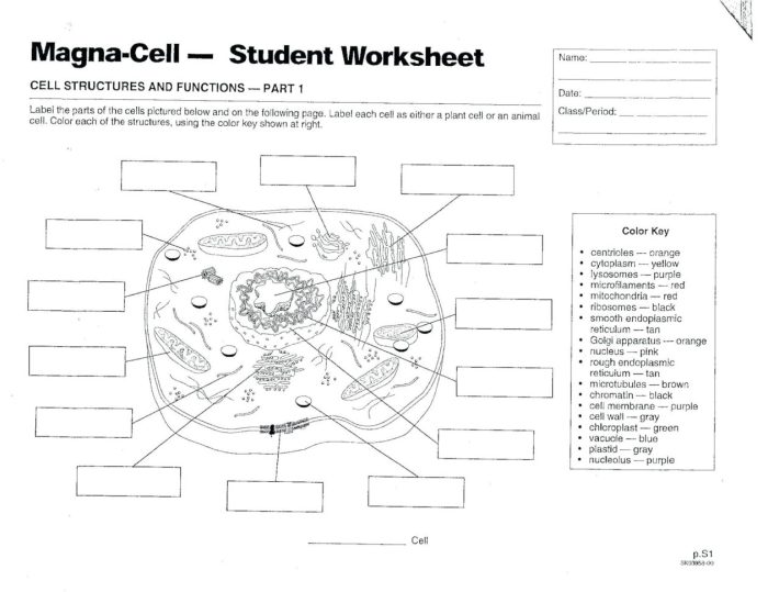
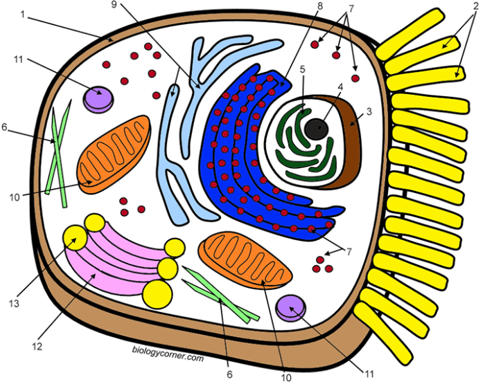
Imagine a simplified, two-dimensional representation of an animal cell. The nucleus, a large, round structure, sits near the center. Scattered throughout the cytoplasm (the jelly-like substance filling the cell) are numerous smaller, oval-shaped mitochondria. The endoplasmic reticulum, a complex network of interconnected membranes, extends throughout the cytoplasm, often appearing as a series of interconnected tubes and sacs.
The Golgi apparatus, resembling a stack of flattened sacs, is typically located near the nucleus. Finally, the cell is enclosed by the plasma membrane, a thin, flexible outer boundary. The cytoskeleton, though not readily visible, provides internal structural support. Ribosomes are too small to be easily depicted in a simplified diagram, but are present throughout the cytoplasm.
Lysosomes are small, membrane-bound vesicles scattered within the cytoplasm.
Differences Between Plant and Animal Cells
Plant and animal cells share some similarities, but also possess key differences. Plant cells possess a rigid cell wall made of cellulose, providing structural support and protection, absent in animal cells. Furthermore, plant cells contain chloroplasts, organelles responsible for photosynthesis, enabling them to produce their own food, a capability animal cells lack. Plant cells typically have a large central vacuole for storing water and other substances, whereas animal cells may have smaller vacuoles or none at all.
These differences reflect the distinct needs and functions of these two types of eukaryotic cells. The presence or absence of these features significantly impacts the overall structure and functionality of the respective cells.
Worksheet Analysis: Animal Cell Coloring Worksheet Answers
Unlocking the secrets of the animal cell is an exciting journey! This section delves into the process of identifying the various organelles within a typical animal cell coloring worksheet, providing a deeper understanding of their roles and the potential challenges students might encounter. By carefully examining the visual representations, students can develop a strong foundation in cell biology.
Analyzing a cell coloring worksheet involves more than just filling in colors; it’s about recognizing the unique shapes and positions of each organelle and connecting them to their specific functions within the cell. This active learning approach transforms a simple coloring exercise into a powerful tool for understanding complex biological structures.
The meticulous detail required for accurate animal cell coloring worksheet answers, with their intricate organelles, sometimes feels strangely akin to the pixelated precision needed for crafting minecraft animals coloring pages. Both demand patience and attention to detail, though one involves the microscopic world and the other, the blocky realm of digital creatures. Ultimately, both activities offer a quiet satisfaction in the completion of a carefully rendered image.
Organelle Identification and Function
The following table summarizes the key features and functions of organelles commonly found in animal cell coloring worksheets. Understanding these features will greatly aid in accurate identification and comprehension of cellular processes.
| Organelle Name | Function |
|---|---|
| Nucleus | Contains the cell’s genetic material (DNA) and controls cell activities. Think of it as the cell’s “control center.” |
| Cytoplasm | The jelly-like substance filling the cell; it suspends the organelles and is the site of many metabolic reactions. Imagine it as the cell’s “workspace.” |
| Cell Membrane | The outer boundary of the cell; it regulates what enters and exits the cell, maintaining its internal environment. It acts like the cell’s “gatekeeper.” |
| Mitochondria | The “powerhouses” of the cell; they generate energy (ATP) through cellular respiration. They are essential for the cell’s energy needs. |
| Ribosomes | Small structures responsible for protein synthesis; they translate genetic information into functional proteins. They are the cell’s “protein factories.” |
| Endoplasmic Reticulum (ER) | A network of membranes involved in protein and lipid synthesis and transport. The rough ER (with ribosomes) synthesizes proteins, while the smooth ER synthesizes lipids and detoxifies substances. |
| Golgi Apparatus (Golgi Body) | Processes, packages, and transports proteins and lipids. It acts as the cell’s “shipping and receiving department.” |
| Lysosomes | Contain digestive enzymes that break down waste materials and cellular debris. They are the cell’s “recycling centers.” |
| Vacuoles | Storage sacs for water, nutrients, and waste products; animal cells typically have smaller, more numerous vacuoles than plant cells. |
Challenges in Organelle Identification
Visual representations on worksheets can sometimes present challenges for students. For instance, the size and shape of organelles may not always be accurately depicted to scale, leading to potential misidentification. Overlapping organelles can also make distinguishing between them difficult. Furthermore, the lack of three-dimensionality in a 2D worksheet can make it challenging to visualize the spatial relationships between different organelles.
For example, distinguishing between the rough and smooth endoplasmic reticulum solely based on a 2D image can be difficult without prior knowledge of their structural differences. Students might also struggle with recognizing the subtle differences in the appearance of organelles like lysosomes and vacuoles, which can look similar in simplified diagrams. Providing multiple examples and varied representations of organelles can help students overcome these challenges.
Coloring Worksheet Design & Pedagogy

Coloring worksheets offer a vibrant and engaging approach to learning about complex biological structures like animal cells. They transform the often-daunting task of memorizing organelles into a fun, hands-on activity that caters to diverse learning styles. The visual nature of the activity enhances comprehension and retention, making it a valuable tool in any science classroom.
This section delves into the design of an effective animal cell coloring worksheet and explores the pedagogical benefits of integrating this technique into a broader lesson plan on cell biology. We’ll examine how strategic color choices and clear labeling can significantly enhance learning outcomes.
Animal Cell Coloring Worksheet Design
The following design incorporates eight key organelles, each assigned a distinct color to aid memorization and visual differentiation. The choice of colors is deliberate, aiming for clarity and visual appeal, avoiding colors that might be too similar or difficult to distinguish. The use of bright, easily identifiable colors is paramount for optimal learning.
- Cell Membrane: Light Blue – Representing the boundary and selective permeability.
- Cytoplasm: Pale Yellow – Highlighting the jelly-like substance filling the cell.
- Nucleus: Dark Purple – Emphasizing its role as the control center.
- Nucleolus: Darker Purple (a shade darker than the nucleus)
-Indicating its function in ribosome production. - Mitochondria: Bright Red – Representing the powerhouse of the cell and energy production.
- Ribosomes: Dark Green – Showcasing their role in protein synthesis.
- Endoplasmic Reticulum (ER): Light Green (a lighter shade than the ribosomes)
– Differentiating between rough and smooth ER is optional but could be achieved with texture variations or a slightly different shade. - Golgi Apparatus: Orange – Representing the packaging and distribution center.
Educational Value of Coloring Worksheets
Coloring worksheets provide a multi-sensory learning experience that strengthens memory and comprehension. The act of coloring engages students actively, fostering a deeper understanding of cell structure than passive learning methods. This hands-on approach is particularly beneficial for visual learners, helping them to internalize the spatial relationships between different organelles within the cell. Furthermore, the labeling aspect encourages students to actively recall and apply their knowledge, reinforcing learning through repetition.
The visual aid serves as a reference point for future study and discussion.
Effective Teaching Strategies Integrating Coloring Worksheets
Effective integration of coloring worksheets requires careful planning and execution. The worksheet shouldn’t stand alone but should be part of a broader lesson plan.
Examples of effective strategies include:
- Pre-coloring Activity: Begin with a brief lecture or interactive discussion on animal cell structure. Then, distribute the coloring worksheet as a reinforcement activity to solidify concepts learned.
- Post-coloring Discussion: After completing the worksheet, engage students in a class discussion about the organelles, their functions, and their relative sizes and locations within the cell. This allows for clarification of any misconceptions and encourages peer learning.
- Worksheet as a Quiz: Use the completed worksheet as a formative assessment tool. Review the completed worksheets, addressing any misunderstandings and providing immediate feedback.
- Differentiated Instruction: Adjust the complexity of the worksheet to cater to different learning levels. For advanced learners, consider adding more organelles or requiring them to research and add information about each organelle’s function.
Interpreting Cell Processes Through Coloring

Coloring a diagram of an animal cell is more than just a fun activity; it’s a powerful tool for understanding complex cellular processes. By actively engaging with the visual representation, students can build a deeper and more intuitive grasp of how different organelles interact and contribute to the cell’s overall function. This active learning approach transforms abstract concepts into tangible, memorable experiences.The coloring activity facilitates understanding of cellular processes by linking visual cues with functional roles.
This direct association strengthens memory and comprehension, making learning more effective and enjoyable. The vibrant colors help to differentiate organelles and their interactions, creating a more engaging and memorable learning experience.
Protein Synthesis Visualization
The process of protein synthesis, from transcription in the nucleus to translation in the ribosomes, can be vividly illustrated using a colored cell diagram.
- The nucleus, colored a deep purple, represents the site of DNA transcription where the genetic code is copied into mRNA.
- The mRNA, depicted in bright yellow, travels from the nucleus (purple) to the ribosomes (light blue).
- Ribosomes, light blue, are shown actively translating the mRNA code, building a polypeptide chain (represented by a string of different colored beads, each bead representing an amino acid).
- The endoplasmic reticulum (ER), colored a pale green, can be shown interacting with the ribosomes, modifying and transporting the newly synthesized protein.
- Finally, the Golgi apparatus (orange), receives and further processes the protein before packaging it into vesicles (small, dark green circles) for transport to other locations within the cell or secretion outside the cell.
Cellular Respiration Spatial Relationships
The spatial arrangement of organelles is crucial for understanding cellular respiration. The colored worksheet provides a clear visual map of these relationships.The visual representation on the worksheet clarifies the flow of molecules and energy during cellular respiration. For example, the proximity of mitochondria (red) to the cytoplasm (pale yellow) highlights the importance of glucose (represented by small, yellow circles) availability for ATP production.
The positioning of the mitochondria near the ER (pale green) illustrates the involvement of the ER in lipid metabolism, which contributes to the production of energy. The relationship between the mitochondria and the nucleus (purple) further reinforces the importance of genetic control in energy production.
Explaining Cellular Processes Using a Colored Worksheet, Animal cell coloring worksheet answers
A student can use the colored worksheet to explain cellular respiration by following these steps:
- Begin by identifying the mitochondria (red) as the primary site of ATP production.
- Trace the pathway of glucose (yellow circles) from the cytoplasm (pale yellow) to the mitochondria.
- Describe the three main stages of cellular respiration: glycolysis (in the cytoplasm), the Krebs cycle (within the mitochondria), and the electron transport chain (in the inner mitochondrial membrane).
- Explain how oxygen (represented by small, blue circles) is crucial for the electron transport chain and ATP synthesis.
- Highlight the production of carbon dioxide (represented by small, grey circles) and water (represented by small, light blue circles) as byproducts of respiration.
- Finally, connect the process to the cell’s overall energy needs and other cellular functions that depend on ATP.


